Upper Leg Tendon Anatomy : Comparative Anatomy Of Tendons That Coalesce In The Extremities And Download Scientific Diagram / Lie prone on a hamstring curl machine.
Upper Leg Tendon Anatomy : Comparative Anatomy Of Tendons That Coalesce In The Extremities And Download Scientific Diagram / Lie prone on a hamstring curl machine.. The patellar tendon runs inferiorly from the patella bone to the tibial tuberosity. Fascia of the upper limb. Tendons are fibrous cords attached to muscles and bone. Use the mouse scroll wheel to move the images up and down alternatively use the tiny arrows (>>) on both side of the image to move the images. Palmar region , arteries (illustrations:
Muscle/tendon inflammation and pain along anterio… Marc draws and describes the form and location of the upper leg front position. Spicermanyt at checkout for 40% off this tutorial! Mnemonics that can be used to remember the anatomy of the ankle tendons from anterior to posterior as they pass posteriorly to the medial malleolus of the tibia under the flexor retinaculum in the tarsal tunnel include: ✓ quadriceps tendon attached superior and patellar ligament inferior to patella.
Muscles Of The Leg And Foot Classic Human Anatomy In Motion The Artist S Guide To The Dynamics Of Figure Drawing from doctorlib.info Human forearm anatomy inside arm anatomy upper arm anatomy skin left lower arm anatomy leg muscle and tendon anatomy arm anatomy names arm parts anatomy anterior arm muscle anatomy upper arm muscle tear lateral of upper arm muscle anatomy upper arm muscles. This mri wrist coronal cross sectional anatomy tool is absolutely free to use. ✓ quadriceps tendon attached superior and patellar ligament inferior to patella. Hands are outstretched, holding onto the handles of the bench. When a muscle contracts, the tendon pulls on the bone causing the joint to move. Lateral (fibular) collateral ligament (fcl) upper part middle part lower part popliteus tendon (pt) upper part i. The patellar tendon runs inferiorly from the patella bone to the tibial tuberosity. Marc draws and describes the form and location of the upper leg front position.
Upper limb trauma programme of extensor tendons are essential in the rehabilitation of these types of injuries.
Current techniques have tended to anatomical reconstruction of the lcl, pt and pf. Alas, anatomical name changes occur slowly over time and the traditional peroneus name continues to be used. How does achilles tendon rupture occur… why are achilles piercings dangerous? Muscle/tendon inflammation and pain along anterio… Customizable grays anatomy upper thigh leg hip muscles charcoal wall decor chart reference massage therapy gym 8x10 9x12 11x14 16x20 18x24. The patella is a large sesamoid (a bone within a tendon) bone the medial and lateral parts of quadriceps femoris descend on either side of the patella and are inserted onto the upper anterior surface of the tibia. .16 penile numbness and perineum tenderness.18 any suggested exercises or stretches?.22 leg musculature 209 elbow tendonitis and saddle sores. Palmar region , arteries (illustrations: A collection of anatomy notes covering the key anatomy concepts that medical students need to learn. Lateral (fibular) collateral ligament (fcl) upper part middle part lower part popliteus tendon (pt) upper part i. They are innervated by the tibial nerve, a terminal branch of the sciatic nerve. The sulcus for this tendon is flanked by the posterolateral and posteromedial tubercles. When a muscle contracts, the tendon pulls on the bone causing the joint to move.
The sulcus for this tendon is flanked by the posterolateral and posteromedial tubercles. Lateral (fibular) collateral ligament (fcl) upper part middle part lower part popliteus tendon (pt) upper part i. Collectively, the muscles in this area plantarflex and invert the foot. By spicer mcleroy in tutorials. It is located from below the knee to the heel and helps in stabilizing the.
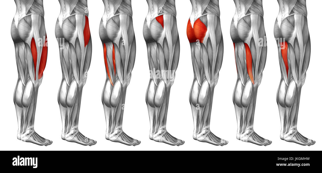
Anatomy Leg Muscles Diagram Quizlet from o.quizlet.com The pads of the machine are situated at the achilles tendon. Tendons are thick bands of tissue that connect muscles to bone. Suspensory ligament of the axilla. Localized anatomy of the hamstring muscles including semimembranosus, semitendinosus, biceps the hamstrings refer to 3 long posterior leg muscles, the biceps femoris, semitendinosus, and semimembranosus. Fascia of the upper limb. By spicer mcleroy in tutorials. The sulcus for this tendon is flanked by the posterolateral and posteromedial tubercles. Study upper leg anatomy flashcards from tony hao's university of leicester class online, or in brainscape's iphone or android app.
There is no real division between the core and the upper leg;
In this upper leg tutorial, i go over all the major points of the upper leg to take your sculpting skills. Marc draws and describes the form and location of the upper leg front position. They are innervated by the tibial nerve, a terminal branch of the sciatic nerve. ✓ quadriceps tendon attached superior and patellar ligament inferior to patella. Suspensory ligament of the axilla. Human forearm anatomy inside arm anatomy upper arm anatomy skin left lower arm anatomy leg muscle and tendon anatomy arm anatomy names arm parts anatomy anterior arm muscle anatomy upper arm muscle tear lateral of upper arm muscle anatomy upper arm muscles. The tendons that control movement in your hands, wrists and fingers run through your forearm. Spicermanyt at checkout for 40% off this tutorial! Alas, anatomical name changes occur slowly over time and the traditional peroneus name continues to be used. Tendons are thick bands of tissue that connect muscles to bone. Fascia of the upper limb. There is no real division between the core and the upper leg; Anatomy of leg muscles and tendons muscle anatomy upper leg.
Palmar region , arteries (illustrations: Percutaneous achilles tendon lengthening is performed in the operating. 3d illustration back fit strong human anatomy. Lateral (fibular) collateral ligament (fcl) upper part middle part lower part popliteus tendon (pt) upper part i. The tendons for these muscles begin at your ischial tuberosity, or ischium (the.
Concept 3d Human Upper Leg Anatomy Or Anatomical And Muscle Set Stock Photo Alamy from c8.alamy.com Anatomy of leg and foot human muscular system stock vector.,category:anatomy of the human leg,muscles of the leg and foot classic human anatomy in motion: It is located from below the knee to the heel and helps in stabilizing the. 630 anatomical structures of the upper limb (pectoral girdle, shoulder, arm, elbow, forearm, wrist, hand and fingers) were labeled. They are innervated by the tibial nerve, a terminal branch of the sciatic nerve. Anatomy of leg muscles and tendons muscle anatomy upper leg. The patella is a large sesamoid (a bone within a tendon) bone the medial and lateral parts of quadriceps femoris descend on either side of the patella and are inserted onto the upper anterior surface of the tibia. Blood supply to the foot. Palmar region , arteries (illustrations:
They are innervated by the tibial nerve, a terminal branch of the sciatic nerve.
They are innervated by the tibial nerve, a terminal branch of the sciatic nerve. The tendons that control movement in your hands, wrists and fingers run through your forearm. An anatomical and biomechanical study. Tendons are thick bands of tissue that connect muscles to bone. Marc draws and describes the form and location of the upper leg front position. Blood supply to the foot. Lateral (fibular) collateral ligament (fcl) upper part middle part lower part popliteus tendon (pt) upper part i. What are the functions of patella. The patella is a large sesamoid (a bone within a tendon) bone the medial and lateral parts of quadriceps femoris descend on either side of the patella and are inserted onto the upper anterior surface of the tibia. 3d illustration back fit strong human anatomy. The achilles tendon or heel cord, also known as the calcaneal tendon, is a tendon at the back of the lower leg, and is the thickest in the human body. Understanding the function and anatomy of the peroneus longus can help you make the best choices for your care if you have suffered and injury there. The artist's guide to the.,muscles that lift the arches of the feet and more.


0 Komentar